




Patient L., 74 years old. 27 hours since the onset of the disease. Neurological status on admission: NIHSS 8 (deep paresis in the left arm, moderate, close to deep, paresis in the left leg, paresis of the left VII cranial nerve, mild left hemihypesthesia, clear consciousness). Brain MSCT showed 2 small ischemic foci in the region of the right basal ganglia (Fig. 2), ASPECTS 8 (Alberta Stroke Program Early CT Score)
History of aortic valve replacement, for which the patient regularly takes a vitamin K antagonist. International normalized ratio on admission was 2.5.
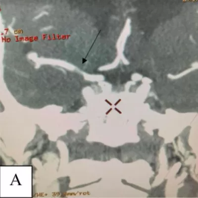
Given the MSCT angiography data (Fig. 1 (A), the angiography data (Fig. 1 (B), and the time since the onset of the disease (27 hours), we initially suspected stenosis (narrowing) of the M1 segment of the right middle cerebral artery . A more detailed examination of the M1 segment after superselective angiography through the Sofia 6F distal access catheter made it possible to verify angiographic signs of thrombus formation at the level of the filling defect (Fig. 1 (C). Given the history of aortic valve replacement and the patient’s use of a vitamin K antagonist with an international normalized ratio of 2.5, embolism (a fibrin-rich thrombus) at the level of the M1 segment of the middle cerebral artery could not be ruled out; it partially occluded the arterial lumen, while the vitamin K antagonist prevented complete occlusion of this arterial segment. In view of the above, we chose a non-standard combination of endovascular tools (the Sofia 6F distal access catheter and the Sceptor C compliant balloon catheter (MicroVention-Terumo)), which made it possible to solve both problems during the operation.
First, the Sceptor C balloon catheter, intended to serve both as a dilation balloon (in case of stenosis) and as a device protecting against distal embolism (in case of an embolus), was prepared and shaped so that its distal part inflated more than the proximal part. This ensured, on the one hand, safe dilation of the stenosis with the proximal part and, on the other hand, anti-embolic protection with the distal part. This type of balloon catheter also allows, if necessary, an intracranial stent to be delivered through its lumen and implanted. The Sofia 6F distal access catheter was positioned 1 mm proximal to the filling defect, and the Sceptor C balloon catheter was positioned with its proximal marker at the level of the proximal edge of the filling defect and 2/3 more distally in the M1 segment. Aspiration was performed simultaneously with balloon inflation.
Angiographic control after completion of the manipulations (Fig. 1 (D) showed complete restoration of patency of the M1 segment of the right middle cerebral artery. Thus, the filling defect of the M1 segment was caused by a fibrin-rich organized thrombus that occluded more than 90% of the arterial segment lumen, and only the patient’s use of a vitamin K antagonist prevented thrombosis of the middle cerebral artery.
After restoration of arterial patency, strength in the left limbs returned within 30 – 40 minutes. By day 3 – complete regression of focal neurological symptoms.


