


Patient B., 7 years old. Preliminary diagnosis: fibrous dysplasia of the left mandible. She was urgently transferred to the SI “SPCENRS of the NAMS of Ukraine” under endotracheal anesthesia from the maxillofacial surgery department with arterial bleeding. The bleeding occurred during open surgery, from the wound, while attempting to remove the dysplasia. The bleeding area was packed.
Due to significant blood loss, a blood transfusion was performed. Immediately upon admission to the Center, the patient was taken to the X-ray operating room. Under endotracheal anesthesia, via transfemoral access, catheterization and angiography of the right and left external carotid arteries (ECA) were performed. The angiogram showed an intact right external carotid artery.
The angiogram of the left ECA shows filling of a hypervascular soft-tissue lesion in the mandibular region. The angiographic picture is that of an arteriovenous malformation (AVM).
The AVM fills from the territories of the alveolar branch of the maxillary artery and from the lingual artery. Moreover, selective arteriography of the lingual artery revealed that the arterial branch to the AVM arises more distally than the branch to the tongue (Fig 1, 2).
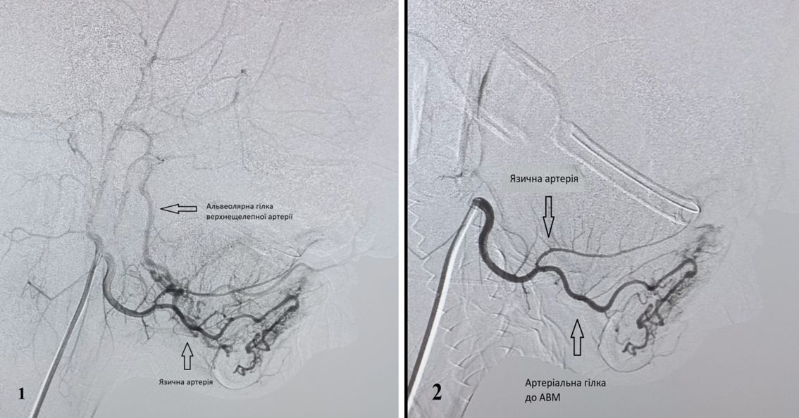
Figure 1,2 – Patient B. Arteriography of the left external carotid artery. The angiogram of the left ECA shows filling of a soft-tissue arteriovenous malformation in the mandibular region. The AVM fills from the territories of the alveolar branch of the maxillary artery and from the lingual artery. The arterial branch to the AVM arises more distally than the branch to the tongue
After assessing the angiographic picture, a decision was made to embolize the AVM in order to stop the bleeding. First, selective catheterization of the alveolar branch of the maxillary artery was performed using a 4F Hinck-type diagnostic catheter over a 0.035 in hydrophilic guidewire, and it was embolized with 125–250 micron PVA emboli through this catheter. The control angiogram shows no filling of the AVM from the territory of the alveolar branch of the maxillary artery (Fig 3, 4).
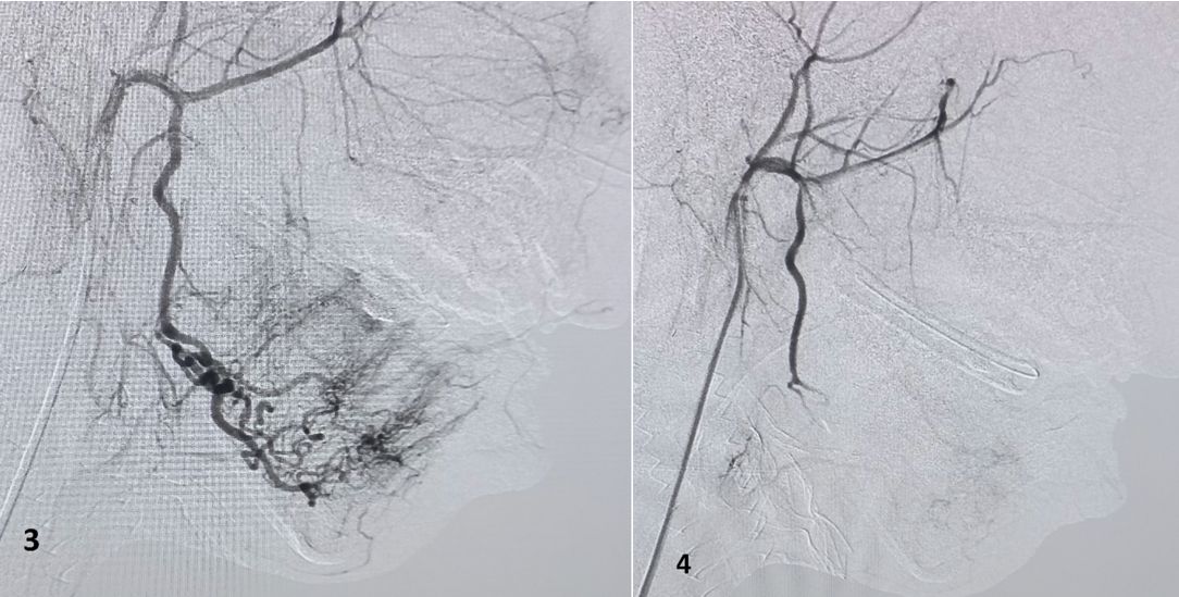
Figure 3, 4 – Patient B. Embolization of the alveolar branch of the maxillary artery. Selective catheterization of the alveolar branch of the maxillary artery and its embolization. The control angiogram shows no filling of the AVM from the territory of the alveolar branch of the maxillary artery.
As a second stage, selective catheterization of the origin of the lingual artery was performed, also using a 4F Hinck-type catheter over a 0.035 in hydrophilic guidewire. However, it proved impossible to advance this catheter further along the lingual artery. Embolization of the AVM from this catheter tip position is impossible due to the high risk of emboli migrating into the lingual artery, which would inevitably lead to ischemia and necrosis of the tongue tissues. Therefore, a decision was made to use a 1.7F neurosurgical microcatheter over a 0.014 in guidewire, which can pass through the inner lumen of the diagnostic catheter. Using the 1.7F microcatheter over a 0.014 in guidewire, it was possible to advance distally along the lingual artery, beyond the origin of the arterial branch to the tongue, and to place the microcatheter tip directly in the artery to the AVM (Fig 5).

Figure 5 – Patient B. Arteriography. The 1.7F microcatheter over a 0.014 in guidewire has been advanced distally along the lingual artery, beyond the origin of the arterial branch to the tongue, and placed directly in the artery to the AVM
Embolization of the AVM was performed through the lumen of the 1.7F microcatheter by injecting 125–250 micron PVA emboli. The control angiogram shows no filling of the AVM from the territory of the lingual artery (Fig 6).

Figure 6 – Patient B. Embolization of the lingual artery branch. The control angiogram shows no filling of the AVM from the territory of the lingual artery.
The operation was completed. The catheters were removed. Hemostasis of the puncture site. Immediately after the operation, the packs were removed from the oral cavity. There is no bleeding from the surgical wound. The patient was extubated and returned to the maxillofacial surgery department.


