


Which arteries supply the brain?
The carotid arteries form the carotid territory. The common carotid arteries originate in the chest cavity: the right one from the brachiocephalic trunk (which arises from the aortic arch and divides into the common carotid and subclavian arteries), the left one from the aortic arch. Approximately in the middle of the neck they divide into external and internal branches. The external carotid arteries supply blood to the soft tissues of the head, and the internal carotid arteries to the brain. The carotid arteries provide about 70-85% of the blood flow to the brain.
The vertebral arteries form the vertebrobasilar territory. They supply the occipital lobes of the brain, the medulla oblongata, the cervical spinal cord, the cerebellum and the subcortical structures of the brain. The vertebral arteries originate in the chest cavity and run to the brain in a bony canal formed by the transverse processes of the cervical vertebrae. According to various data, the vertebral arteries provide about 15-30% of the blood flow to the brain.
The brachiocephalic trunk, the carotid and subclavian arteries together with their first branches (the vertebral arteries, etc.) are also called the brachiocephalic arteries (brachio – shoulder, cephalis – head (Greek), i.e. shoulder-head arteries).
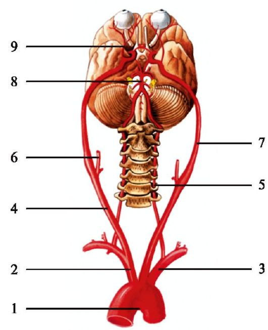
Main arteries of the brain
1 – aortic arch; 2 – brachiocephalic trunk; 3 – left subclavian artery; 4 – right common carotid artery; 5 – vertebral artery; 6 – external carotid artery; 7 – internal carotid artery; 8 – basilar artery; 9 – ophthalmic artery.
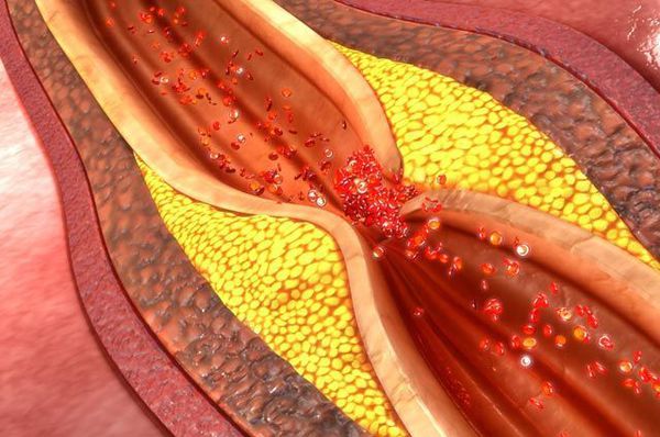
Atherosclerosis is a chronic disease of the arteries caused by impaired lipid and protein metabolism, in which large amounts of cholesterol are deposited in the vascular wall, forming thickenings – atherosclerotic plaques. They gradually narrow the lumen of the arteries and make them less elastic. This obstructs normal blood flow and leads to insufficient blood supply to a particular organ. Narrowing of the vessel lumen by an atherosclerotic plaque is called stenosis, and complete blockage of the lumen is called occlusion. Symptoms of the disease begin to appear at a certain degree of arterial stenosis.
A complicated atherosclerotic plaque forms as the process progresses. There is a significant increase in the lipid core, increased aggregation (sticking together) of platelets, hemorrhages into the plaque, thinning of its fibrous capsule and destruction of the plaque cap with the formation of fissures, ruptures and atherosclerotic ulcers. Thrombi form at the ulcers, and later atherocalcinosis develops (deposition of calcium salts in the atheromatous masses, the plaque structure). The main consequence of a complicated atherosclerotic plaque is the formation of a mural thrombus, which suddenly and sharply restricts blood flow in the artery. Atherosclerosis of the brachiocephalic arteries (BCA) threatens ischemic stroke (cerebral infarction).
What precedes atherosclerosis?
This disease has its own risk factors. Atherosclerosis develops as a consequence of:
- smoking;
- arterial hypertension (systolic BP > 140 mmHg, diastolic BP > 90 mmHg);
- impaired carbohydrate metabolism (hyperglycemia, diabetes mellitus)
- obesity;
- hyperhomocysteinemia;
- dyslipidemia (increased blood levels of certain cholesterol fractions);
- an unhealthy diet (eating large amounts of fats and carbohydrates)
- a sedentary lifestyle (physical inactivity);
- frequent stress;
- postmenopause and hyperfibrinogenemia;
- hereditary predisposition.
Risk factors also include age (people over 50–60 years) and male sex.
Factors such as smoking, physical inactivity and eating habits relate to lifestyle: they can be modified (are correctable). Modifiable risk factors are elevated blood pressure, type 2 diabetes mellitus and dyslipidemia. Age, male sex and heredity are non-modifiable risk factors.
Diagnosis of atherosclerotic pathological changes of the BRACHIOCEPHALIC ARTERIES.
- Ultrasound examination of the head and neck vessels (duplex, triplex scanning)
- Computed tomography (CT) angiography
- Magnetic resonance angiography (MRA)
- Digital subtraction cerebral angiography (DSA)
Ultrasound examination of the brachiocephalic arteries is an excellent non-invasive method that combines high informativeness and low cost for obtaining preliminary information about the degree of vascular damage, the nature of blood flow and the condition of the surrounding tissues. However, it should be noted that an ultrasound procedure does not provide comprehensive information about the condition of a vessel. Establishing indications for surgery (surgical removal of the plaque or endovascular stenting) solely on the basis of ultrasound of the brachiocephalic arteries is unacceptable! Stenoses diagnosed by ultrasound require additional examinations of the vascular system of the neck and head (CT angiography, MRA or DSA). Only when two different examination methods confirm a high degree of stenosis does the surgeon have the right to decide on a particular method of surgical correction of brachiocephalic artery patency.
Laboratory diagnostics should include
- lipid profile (total cholesterol, triglycerides (TG), low-density lipoproteins (LDL), very-low-density lipoproteins (VLDL), high-density lipoproteins (HDL), atherogenic index;
- complete blood count;
- coagulogram;
- blood sugar;
- total blood protein;
- thyroid hormones (T3, free T4);
- creatinine, urea;
- ALT, AST, GGT, bilirubin.
TREATMENT
1. Conservative. Optimal drug therapy is possible if the stenosis at the plaque level is < 60% and has not manifested itself in any way (asymptomatic course):
- there are no ischemic foci on brain MRI or MSCT in the territory of the artery affected by atherosclerosis.
- there has not been a single episode of neurological deficit (transient ischemic attack) corresponding to impaired blood supply to the brain area that receives blood from the territory of the artery stenosed by the atherosclerotic plaque.
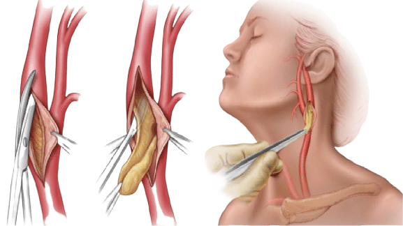
- Absence of characteristic features of a complicated atherosclerotic plaque according to the examinations performed. The presence of atherosclerotic ulcers and signs of plaque cap destruction, even with small stenoses, is an indication for endovascular stenting or carotid endarterectomy (surgical removal of the plaque).
2. Surgical.
The 2017 guidelines of the European Society of Cardiology (ESC) and the European Society for Vascular Surgery (ESVS) clearly set out the indications for surgical correction of stenoses in the section on diseases of the extracranial carotid and vertebral arteries (pp. 182 – 191).
Algorithm for managing patients with disease of the extracranial carotid artery segments

For atherosclerotic lesions of the carotid arteries, two methods of surgical correction are used:
CAROTID ENDARTERECTOMY
ENDOVASCULAR STENTING

The main advantages of the endovascular method are low invasiveness (the operation is performed through an arterial puncture in the upper or lower limb), the possibility of simultaneous stenting (restoring arterial patency by implanting a stent into the vessel lumen) of the extracranial segments of several arteries in any arterial territories (both carotid arteries, or a carotid and a vertebral artery, etc.), and simultaneous stenting of stenoses of arteries inside the skull (intracranial arteries) and extracranial ones when both are affected by atherosclerosis.
TREATMENT EXAMPLES
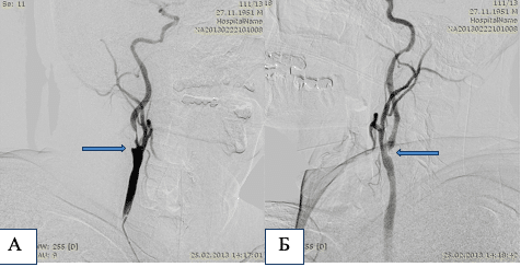
Case No. 1
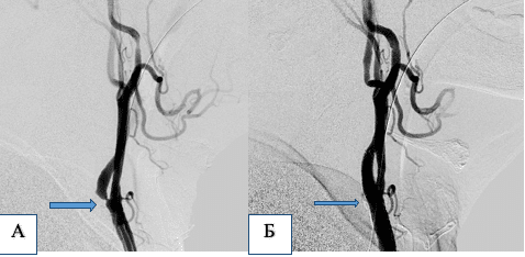
Patient M, 78 years old. Critical stenosis of the left internal carotid artery (ICA).
А – angiogram before the operation. Б – angiogram after endovascular angioplasty and stent implantation at the level of the stenosis.
Case No. 2
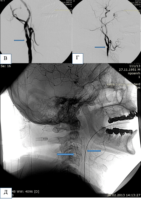
Patient P, 61 years old. Critical stenosis of the right (А) and left (Б) internal carotid arteries.
А and Б – angiograms before the operation. The stenosis sites are marked with arrows. В – angiogram of the right carotid territory after endovascular angioplasty and stent implantation at the level of the stenosis. Г – angiogram of the left carotid territory after endovascular angioplasty and stent implantation at the level of the stenosis. Д – native image of the bifurcation levels of the right and left common carotid arteries showing the implanted stents at the levels of the stenoses of both internal carotid arteries.
Case No. 3
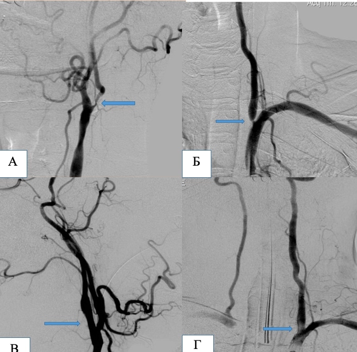
Patient A, 63 years old. Critical stenosis of the left internal carotid artery (А) and the left vertebral artery (Б).
А and Б – angiograms before the operation. The stenosis sites are marked with arrows. В – angiogram of the left carotid territory after endovascular angioplasty and stent implantation at the level of the stenosis. Г – angiogram of the left subclavian artery after endovascular angioplasty and stent implantation at the level of the stenosis of the left vertebral artery ostium.
Case No. 4
Tandem (intra-extracranial) stenoses
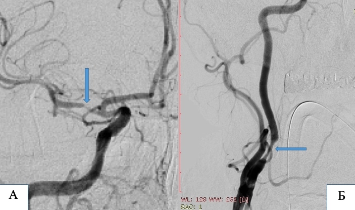
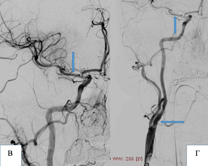
Patient S, 65 years old. Critical stenosis of the M1 segment of the right middle cerebral artery (А) and the right internal carotid artery (Б).
А and Б – angiograms before the operation. The stenosis sites are marked with arrows. В – angiogram of the right carotid territory. The arrow marks the M1 segment of the right middle cerebral artery after implantation of an intracranial stent. The stenosis has been completely eliminated and arterial patency restored. Г – angiogram of the right carotid territory after stenting of the tandem stenoses of the right middle cerebral artery and the right internal carotid artery.
Case No. 5
Intracranial stenosis
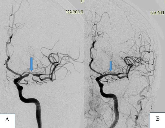
Patient G, 65 years old. Critical stenosis of the M1 segment of the left middle cerebral artery.
А – angiogram before the operation. The stenosis site is marked with an arrow. Б – angiogram of the left carotid territory. The arrow marks the M1 segment of the left middle cerebral artery after implantation of an intracranial stent. The stenosis has been completely eliminated and arterial patency restored.
Case No. 6
Intracranial stenosis
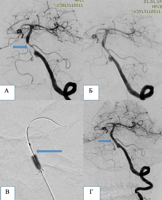
Patient S, 68 years old. Critical stenosis of the proximal segment of the basilar artery.
А – angiogram before the operation. The stenosis site is marked with an arrow. Б – angiogram of the vertebrobasilar territory. Positioning of the balloon-mounted stent by its markers relative to the stenosis site. В – Stent implantation by inflation of the balloon. Г – The arrow marks the proximal segment of the basilar artery after stent implantation. The stenosis has been completely eliminated and arterial patency restored.