


Ultrasound diagnostic
Ultrasound diagnostics (ultrasound), as you know, is sound or acoustic waves, the frequency of which is higher than the maximum frequency of sound, perceives the human ear, namely above 20 kHz. Mostly in ultrasound, a frequency from 2.5 to 12-15 MHz is used. Ultrasound is based on receiving echoes from structures with different densities. Different tissues of the body (muscles, blood, liver, etc.) have different densities, therefore, the propagation of ultrasound in them is also different. On this difference, using the physical properties of ultrasound as a wave, the ultrasound method is based. The ultrasonic waves come from the transducers, which also receive the echoes. The echo signals of the transducer are converted into pulses and displayed on the screen of the device.
So, ultrasound is one of the most effective and non-invasive methods in modern medical diagnostics.
Equipment:
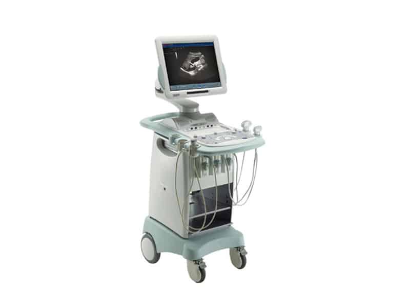
Ultrasonic scanner Еsaote MyLab 40 CV, phased, convection, linear sensors. Pulsed-wave, constant-wave Doppler, modes of color Doppler coding speed, intensity of blood flow. Doppler imaging of tissues, the possibility of synchronization with ECG. A complete package of calculations for cardiological and vascular diseases (echocardiography, vessels of the neck, upper and lower limbs), synchronization with the ECG. Possibility of ultrasound examination of abdominal organs, musculoskeletal system, thyroid, mammary gland. Perform a transcranial triplex scan.
Echocardiography
Echocardiography –The method of ultrasound diagnostics aimed at the study of morphological and functional changes in the heart, valve apparatus, and main vessels.
Indications for Echocardiography are any symptoms that may indicate cardiopathology. Echocardiography is also performed in patients after acute cerebral circulation disorder with suspicion of a cardioembolic subtype of ischemic stroke (search for a cause). This diagnostic method allows you to identify and monitor the congenital and acquired heart valve defects, diagnose additional shunts, septal defects, diagnose cardiomyopathy, detect heart changes and complications in hypertensive disease, coronary heart disease. This method is shown as control after heart and major vessel surgery, deep vein thrombosis, chemotherapy in oncology, suspicion of aortic aneurysm. Also, the indication for Echocardiography is a chronic headache, the presence of hereditary diseases in relatives of the first line (for example, hypertrophic cardiomyopathy and many others.) An example of visualization of valvular pathology, in the comparison format: stenosis – norm