


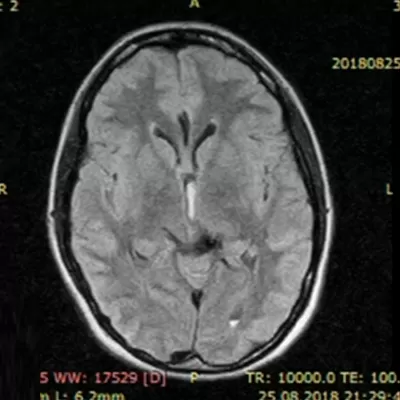
Patient Kh, 31 years old, was admitted to the Center on 03.01.2019 with complaints of periodic headaches. She was transferred to the Center from the SI “Institute of Pediatrics, Obstetrics and Gynecology named after Academician O.M. Lukyanova of the NAMS of Ukraine”, where she had given birth by caesarean section 10 days earlier. No focal neurological symptoms were found at the time of admission.
Her history shows that she fell ill suddenly on 25.08.18, at 20 weeks of pregnancy. A severe headache, nausea and vomiting appeared. She was hospitalized at her place of residence. After brain MRI (26.08.18), rupture of an arteriovenous malformation (AVM) in the region of the posterior third of the corpus callosum, the left lateral ventricle and the left parietal lobe of the brain with intraventricular hemorrhage was diagnosed (Fig. 1). For further treatment, the patient was transferred to the SI “Romodanov Neurosurgery Institute of the NAMS of Ukraine”, where a comprehensive examination was performed (brain MSCT, cerebral angiography). In addition to the AVM, multiple cerebral saccular aneurysms (SA) were diagnosed: SA of the anterior cerebral – anterior communicating artery (ACA-ACoA) on the left, SA of the ACA (A2-A3 segment) on the right and SA of the ACA (A3-A4 segment) on the right. Together with the obstetrician-gynecologists of the SI “Institute of Pediatrics, Obstetrics and Gynecology named after Academician O.M. Lukyanova of the NAMS of Ukraine”, a conservative treatment strategy was chosen, given the high risk of surgery for the mother and fetus, with subsequent surgical treatment after delivery.
On 05.10.18, at 27 weeks of pregnancy, the patient had a recurrent intraventricular hemorrhage, confirmed by brain MSCT on 05.10.18 (Fig. 2). Given the high risk of repeated AVM rupture, the SI “Romodanov Neurosurgery Institute of the NAMS of Ukraine” recommended endovascular surgery aimed at embolization of the AVM nidus; however, the patient and her husband preferred conservative treatment with subsequent surgical treatment after delivery. Conservative therapy was carried out. She was discharged for outpatient treatment at her place of residence with recommendations to deliver at the SI “Institute of Pediatrics, Obstetrics and Gynecology named after Academician O.M. Lukyanova of the NAMS of Ukraine”.
In our Center (after delivery), selective cerebral angiography was performed, and the angioarchitecture of the AVM and the radiological anatomy of the cerebral saccular aneurysms were clarified (Fig. 3). On 10.01.19, endovascular surgery was performed: subtotal embolization of the AVM and endovascular occlusion of the SA of the ACA (A2 – A3 segment) on the right (Fig. 4).
The patient was admitted to the Center for a follow-up examination on 09.04.19. No focal neurological symptoms were found at the time of admission. According to selective cerebral angiography (09.04.19), the AVM is totally excluded from the circulation. The SA of the ACA (A2 – A3 segment) on the right is totally and reconstructively excluded. The aneurysms (ACA-ACoA) on the left and the SA of the ACA (A3-A4 segment) on the right, which had been detected earlier, are not visualized during this examination – normalization of the angiographic picture of the cerebral arteries after total embolization of the AVM due to elimination of the hemodynamic load on the arterial walls caused by pathological arteriovenous shunting (Fig. 5).


