A case of combined (microsurgical and endovascular) treatment of brain arteriovenous malformations
Treatment of cerebral AVMs remains one of the greatest challenges in cerebrovascular neurosurgery. The advantages of surgery are radical removal of the AVM in most cases and a low or practically absent risk of hemorrhage after removal.Preliminary data indicate favorable clinical outcomes of combined treatment with careful patient selection. Preoperative embolization is safe and reduces the size of the AVM and the risk of rupture in malformations with associated aneurysms or high-flow shunts .
Over the past decade, all methods of treating this pathology have evolved. Today, AVMs that were previously considered incurable are removed safely. Microsurgical removal remains the best treatment option in selected patients with AVMs without risk of neurological deterioration, owing to the low risk of recurrence and the absence of a latent period before hemorrhage. A significant improvement in AVM treatment outcomes has been reported in patients with preoperative embolization, which helps reduce blood flow in the AVM, especially in deep feeding vessels, facilitating surgical removal of the malformation.
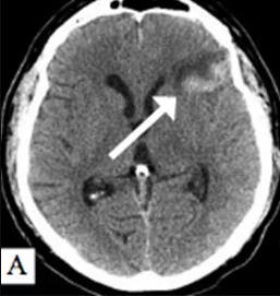
Patient A., born 1976. Arteriovenous malformation of the left temporal lobe of the brain, associated saccular aneurysms of the M1-M2 segment of the left middle cerebral artery and of the cavernous segment of the left internal carotid artery: A – brain computed tomography after the onset of severe headache and speech disorders (subarachnoid-parenchymal hemorrhage with a stroke-hematoma of the frontal lobe). History of generalized epileptic seizures since 1994; B, C – cerebral angiography: a saccular aneurysm of the M1-M2 segment of the left middle cerebral artery with a wide neck and a diverticulum at the dome, and a saccular aneurysm of the cavernous segment of the left internal carotid artery were found. Arteriovenous malformation of the left temporal lobe measuring 6.8 × 4.8 mm, of angiomatous structure, fed by numerous branches of the middle cerebral, internal carotid and posterior cerebral arteries, with superficial and deep venous drainage. A single-stage surgical intervention was performed: presurgical partial endovascular embolization of the arteriovenous malformation (D), clipping of the saccular aneurysm of the left middle cerebral artery and microsurgical removal of the arteriovenous malformation of the left temporal lobe (E, F)
Need a specialist consultation? Leave a request and we will contact you.