


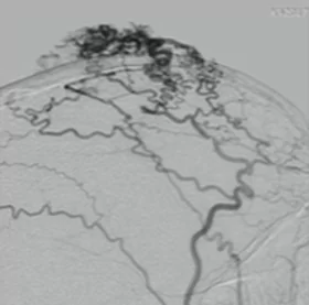
H.P. Krayenbuhl and G. Yasargil, in a review of 800 cases of arteriovenous malformations (AVMs) from the literature and their own clinical experience, found that extracranial AVMs account for only 8.1 % of cases. AVMs of the scalp soft tissues are rare abnormal fistulas between feeding arteries and draining veins without a capillary bed in the subcutaneous-aponeurotic layer. They are rare compared with other subcutaneous or craniofacial vascular anomalies such as hemangioma or venous malformations. N.D. Fisher-Jeffes et al. reported that 38 % (9 of 24) of these malformations were associated with trauma. Traumatic AVMs of the scalp soft tissues usually develop within the superficial temporal artery and present as a scalp swelling that gradually increases in size. Symptoms may include headache, fainting, tinnitus, and rarely bleeding.
The superficial temporal artery is prone to injury because of its superficial course over the temporal bone and its proximity to the cranial sutures. Besides traumatic etiology, arteriovenous fistulas (AVFs) of the superficial temporal artery may also be spontaneous or occur after surgical procedures such as ventriculostomy or craniotomy.
Two mechanisms of formation of traumatic scalp AVFs are proposed. The first is simultaneous rupture of the artery and an adjacent vein, which directly leads to fistula formation; the second is rupture of the vasa vasorum in the arterial wall and proliferation of endothelial cells from the damaged vasa vasorum, which subsequently forms numerous small vessels creating a fistula between arteries and veins.
Diagnosis of a traumatic fistula of the superficial temporal artery is based on the history of trauma and a detailed physical examination. Although angiotomography can visualize the fistula quickly, digital subtraction angiography remains the gold standard of examination, making it possible to identify feeder vessels and draining veins, exclude intracranial components, and obtain information on the direction and velocity of blood flow. Some traumatic fistulas of the superficial temporal artery may not pulsate or may pulsate due to wave transmission from adjacent arteries, which indicates a pseudoaneurysm, true aneurysm, cyst, abscess, hematoma or meningeal artery aneurysm with bone erosion. All of the above are part of the differential diagnosis of a traumatic AVF of the superficial temporal artery.
When treating an AVF of the superficial temporal artery, the high blood flow through the fistula, complex vascular anatomy and the cosmetic problem should be taken into account. Treatment is aimed at reducing the cosmetic defect, preventing bleeding and ischemic skin erosion, and reducing the symptoms of the disease, headache and tinnitus. Possible treatment options: surgical excision, vessel ligation, transarterial and transvenous embolization, injection of sclerosing agents into the fistula.
Endovascular embolization has a whole arsenal of tools and can be used as monotherapy or as an adjuvant to surgical removal, reducing perioperative risks and blood loss during removal of the fistula, especially with large fistulas. Surgical removal of the fistula with ligation of the feeding vessels remains the method of choice.
Example No. 1 Treatment of a patient with an arteriovenous fistula: A — preoperative photo showing the fistula, hypertrophied vessels of the cranial vault and local trophic skin changes over the fistula; B — preoperative angiography; C, D, E — result of endovascular embolization of the fistula; F — result of surgical removal of the fistula.


