


Fusiform aneurysms (FA) account for 15 % of all arterial aneurysms . According to other data, FA occur in 3–13 % of intracranial aneurysms and are located predominantly (in 72–76 % of cases) in the vertebrobasilar territory (VBT) . FA of the basilar artery (BA) account for 36.5 % of all FA and over 78 % of VBT FA .Regardless of the cause, 4 characteristic stages are distinguished in the formation of fusiform aneurysms: fragmentation of the internal elastic lamina and thickening of the intima of a certain segment of the vessel, neoangiogenesis in the thickened intima and thinning of the internal elastic layer, formation of a hematoma in the vessel wall with subsequent thrombosis, recanalization and organization of the thrombus with formation of emboli or hemorrhage.Depending on the stage of pathogenesis, BA FA may have diverse clinical manifestations that are “atypical” for arterial aneurysms. In 1.3 % of cases FA are asymptomatic.
In Japan, the incidence of FA is 7.7 % among patients with stroke: 6.4 % among those with ischemic stroke and 12.1 % among those with hemorrhagic stroke . In 31 % of cases BA FA caused obstructive hydrocephalus and manifested as palsy of the trigeminal, oculomotor, facial and auditory nerves.
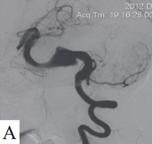
Fusiform aneurysm of the basilar artery. Cerebral angiography: A — before surgery; B — LEO PLUS stent in the lumen of the basilar artery; C — follow-up examination 6 months after surgery


