


Uterine fibroids (fibromyoma, leiomyoma) — one of the most common tumors in women of reproductive age. Externally, uterine fibroids are nodes ranging in size from a pea to a newborn’s head, located in the muscular wall of the uterus (fig. 1). The presence of fibroids in the uterus causes the uterus to enlarge and its shape to become deformed. Therefore, the size of fibroids is usually expressed in “weeks of pregnancy” (for example: uterine fibroid of 9 weeks)
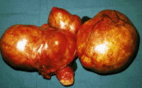 Fig.1
The main symptoms of uterine fibroids are heavy periods, uterine bleeding, pain, and a feeling of heaviness and pressure in the lower abdomen. In 30% of women the development of uterine fibroids is not accompanied by pronounced clinical manifestations, and the tumor may be detected only during a routine gynecological examination or found incidentally during an ultrasound examination.
Treatment: Uterine fibroids can be treated in different ways. With small nodes, drug treatment is possible, consisting of two stages: induction, which reduces the size of the fibroids as much as possible, and stabilizing, which restrains further growth of the fibroid until menopause. With large nodes, surgery is most often used. There are several types of operations: Hysterectomy – complete removal of the uterus together with the nodes. Myomectomy – removal of the fibroid node while preserving the uterus. Uterine artery embolization – a minimally invasive organ-preserving method, without incisions or general anesthesia. The technique consists of inserting a catheter into the femoral artery under local anesthesia. Then a thin microcatheter is advanced into the lumen of the uterine artery. Then small particles (emboli) are injected through the catheter until the vessels feeding the fibromyomatous nodes are blocked. This results in the death (infarction) of these nodes and their subsequent shrinkage (Fig. 2,3).

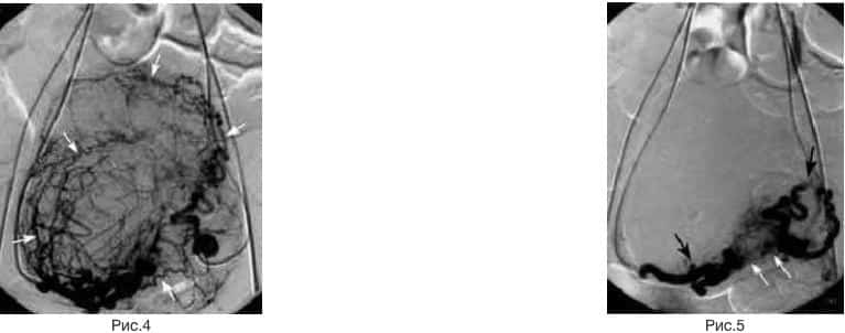
Example: A patient with a large uterine fibroid (12 weeks). Figure 4 shows filling of the fibroid vessels through catheters inserted into the uterine arteries. The borders of the fibroid are marked with arrows. Figure 5 shows the condition after emboli were injected into the fibroid vessels. Only the trunks of the uterine arteries are visible in the image. The fibroid vessels are not visible. They are blocked by emboli.
On the first day after the procedure, embolization is accompanied by severe pain, which requires a hospital stay for pain-relief therapy. Overall, the severity of fibroid symptoms decreases in approximately 85% – 93% of patients. The mortality of the procedure is practically zero, and complications are extremely rare. Successful pregnancies have been reported after selective bilateral uterine artery embolization.
Varicocele – varicose dilation of the spermatic (testicular) vein and the venous plexus surrounding the left testicle. Varicocele occurs with a frequency of 10-16% and is found in every fifth adolescent aged 14-15 years. The development of varicocele is associated with impaired blood outflow due to incompetence of the testicular vein valves. Clinically, it manifests as varicose nodes in the scrotum that are visible or palpable (Fig 1). Dull, aching pain in the groin and testicle, especially after physical exertion.
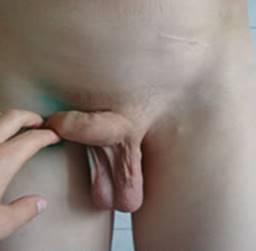 Fig.1
Pronounced varicose dilation of the spermatic vein is accompanied by impaired gonadal function and is a leading cause of infertility. Varicocele is associated with 30-50% of childless marriages.
Several types of surgical correction of varicose gonadal veins have been proposed (Ivanissevich, Bernardi and Palomo operations) and more than 20 of their modifications. The Ivanissevich operation has the largest number of supporters. The operation consists of ligation and transection of the testicular vein in the retroperitoneal space at the level of the iliac vessels. Rational surgery leads to the disappearance of varicocele in 87-97% of patients. However, recurrences occur in 10-87.2% of cases, averaging 22.9%.
As an alternative to the classic Ivanissevich and Palomo operations, methods of X-ray endovascular embolization of the spermatic vein are being actively introduced into surgical practice.
Advantages: minimal invasiveness, high efficiency, safety and technical simplicity. The efficiency of X-ray surgical operations is 87-95.5%. The recurrence rate ranges from 2 to 6%. Spermatic vein embolization is a one-day operation. One day later the patient returns to a normal lifestyle.
Spermatic vein embolization is performed by puncturing the right common femoral vein and successively catheterizing the left renal and spermatic veins, followed by occlusion of the spermatic vein lumen with a coil and injection of a sclerosing solution that seals the vein lumen Fig 2.
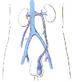 Fig.2
Example: A patient with grade 3 varicocele. The catheter was advanced into the left renal and left spermatic vein. The image shows a sharply dilated left spermatic vein. Reflux of contrast agent into the scrotal veins is noted (Fig. 3.). Embolization of the left spermatic vein was performed by placing an embolization coil (arrow) into its lumen Fig 4.

Benign prostatic hyperplasia (BPH) – (formerly called prostate adenoma). Prostate adenoma is a benign tumor that develops from the glandular epithelium or the stromal component of the prostate, forming nodules that gradually compress the urethra (Fig. 1).
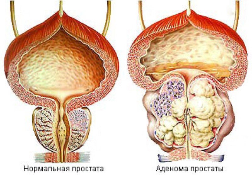 Fig.1
Benign prostatic hyperplasia (BPH) is the most common urological disease in men. According to statistics, after the age of 40 this disease is present in 5-8% of men. As many as 50% of men aged 50 – 60 consult a doctor about BPH. At the age of 70 – 89 it develops in 85% of men.
Depending on the severity of the disease, the main complaints are related to impaired urination due to compression of the urethra by the enlarged prostate. It manifests as a weak urine stream, frequent urges and frequent urination, a feeling of incomplete bladder emptying, urination in small portions, involuntary urination, acute urinary retention, and symptoms of chronic renal failure join in
Treatment: Traditional surgical treatment – complete removal of the prostate. Transurethral resection – removal of the part of the prostate that protrudes into the lumen of the urethra. Prostatic artery embolization for benign prostatic hyperplasia is a new innovative direction in interventional radiology. The technique consists of inserting a catheter into the femoral artery under local anesthesia. Then the catheter is advanced into the lumen of the pelvic arteries. An image of the pelvic vessels is taken to identify the prostatic arteries (Fig 2.)
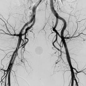 Fig.2
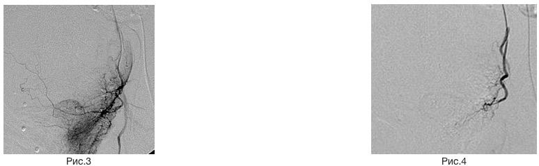
Then microcatheters are used to enter the prostatic arteries. Then small particles (emboli) are injected through the microcatheter until the vessels feeding the prostate adenoma are blocked. This results in the death (infarction) of the adenoma and its subsequent shrinkage, which frees the lumen of the urethra. (Fig. 3, 4).
Example. Computed tomography of a prostate adenoma before and 6 months after embolization. The volume of the prostate decreased by half (Fig. 5.).
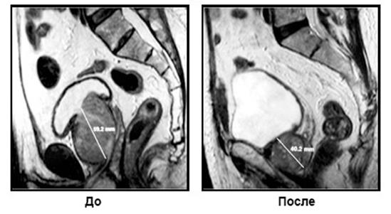 Fig.5
Arteriovenous malformations (angiodysplasias) and hemangiomas are congenital anomalies of the vascular system caused by impaired embryonic morphogenesis, which manifest as hyper-, hypo- and aplasia of arteries, veins, arteriovenous fistulas, capillaries and lymphatic vessels and lead to various forms of impaired regional blood circulation.
The severity of clinical manifestations, significant anatomical and functional disorders, and early disability of patients with vascular malformations of various locations make the problem relevant and call for in-depth study of the diagnosis and treatment of this pathology (Fig 1,2,3).
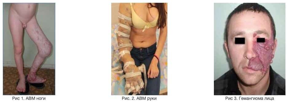
The location of AVMs and hemangiomas is extremely diverse. AVMs and hemangiomas can occur anywhere in the body where there are vessels
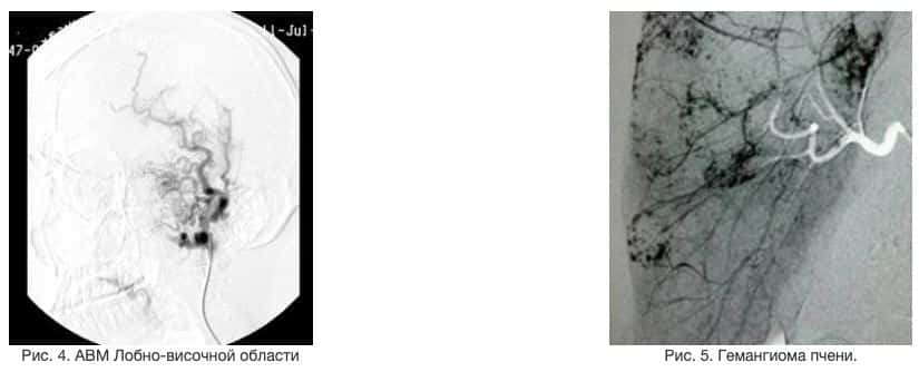
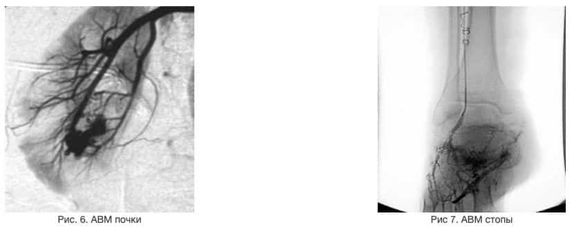
Embolization of AVMs and hemangiomas as a standalone treatment is used:
- in urgent situations to stop bleeding;
- when the AVM or hemangioma is located in areas that are hard to access surgically;
- when other clinical manifestations prevail over cosmetic ones;
- when surgical treatment is declined.
Example 1. Pulmonary AVM. Patient Ya., 19 years old. Complaints of moderate shortness of breath at rest, which worsens after physical exertion. Shortness of breath since childhood. In December 2016 he was admitted to the regional hospital at his place of residence with suspected pneumonia. The chest X-ray showed a shadow in the middle third of the left lung. A chest CT was performed. A diagnosis of pulmonary AVM was made (Fig. 1,2). Pulmonary angiography was recommended. At the SI “SPCENRS of the NAMSU” pulmonary angiography was performed on 01.02.17. Multiple A-V shunts between branches of the pulmonary artery and the pulmonary veins in the lower lobes of the lungs were found, more pronounced on the left

Fig. 1,2. Chest CT. An AVM is seen in the lower lobe of the left lung.
For treatment, on 01.02.17 the patient underwent embolization of 4 large arteriovenous shunts of the left lung with Cook “Tornado” embolization coils. (Fig. 3,4). The operation was technically successful. The postoperative period was uneventful. Improvement was noted in the form of disappearance of shortness of breath at rest. Discharged in satisfactory condition under the supervision of a vascular surgeon and a pulmonologist at the place of residence.

Example 2. Renal AVM. Patient K., 27 years old. Complaints of hematuria. A diagnosis of AVM of the left kidney was made. At the SI “SPCENRS of the NAMSU” renal angiography was performed on 23.05.17. A-V shunts in the lower pole of the left kidney were found (Fig. 1,2,3.).
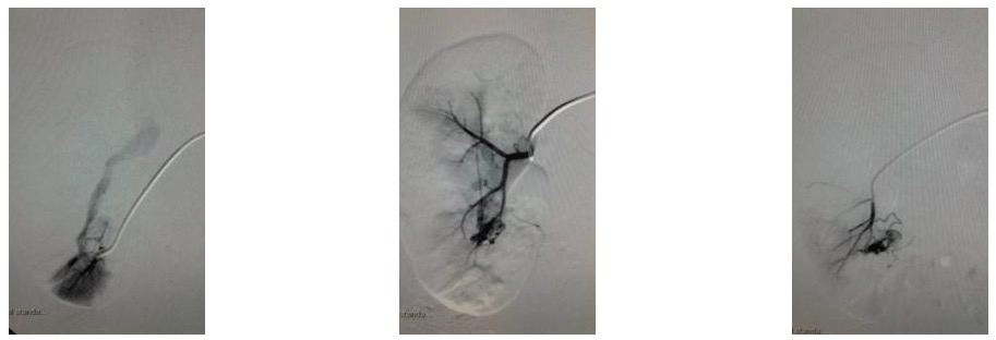
Fig 1,2,3. AVM of the left kidney.
For treatment, on 23.05.17 the patient underwent embolization of the arteriovenous shunts of the left kidney with 400 – 700 micron emboli. (Fig. 5,6). The operation was technically successful. The postoperative period was uneventful. The hematuria stopped. Discharged in satisfactory condition under the supervision of a vascular surgeon and a urologist at the place of residence.

Fig 5,6. Embolization of the left kidney AVM. The follow-up angiogram shows no filling of the AVM.
Example 3. Hemangioma of the lower limb. Patient P., 11 years old. Complaints of a dense tumor-like lesion within the muscle tissue of the lower third of the right lower leg, and pain in the calf muscles during physical exertion. Examination at the place of residence and at the “OKHMATDYT” clinic, based on CT, MRI and Doppler ultrasound data, led to a diagnosis of a hypervascular tumor of the right lower leg. At the SI “SPCENRS of the NAMSU”, for diagnostic purposes, angiography of the right lower limb was performed on 06.10.16. A hypervascular tumor located within the muscles of the right lower leg, probably a hemangioma, was diagnosed. (Fig. 1,2.). To clarify the diagnosis, a puncture biopsy of the tumor was performed at the Institute of Oncology. Histology confirmed the diagnosis of hemangioma.
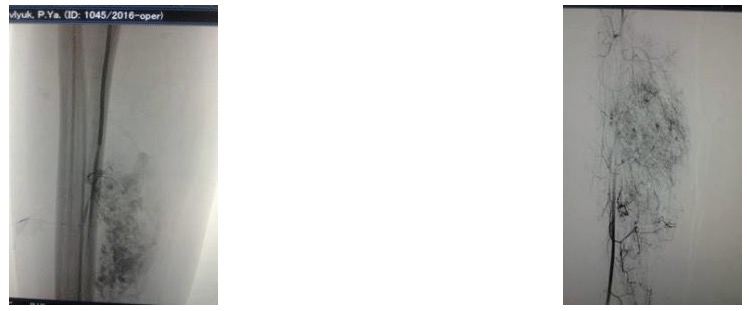
Fig. 1,2. Hemangioma of the right lower leg
For treatment, on 19.10.16 endovascular embolization of the hemangioma was performed with 300 micron microemboli in the territory of the peroneal and anterior tibial arteries by superselective microcatheterization. (Fig.3,4,5,6,7). In the postoperative period, anti-inflammatory, analgesic and antibiotic therapy was given. Discharged in satisfactory condition under the supervision of a vascular surgeon at the place of residence
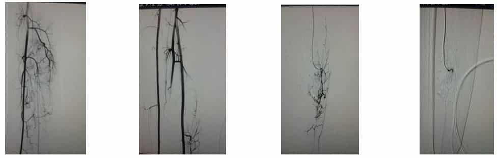
Fig. 3,4,5,6. Stages of embolization of the right lower leg hemangioma
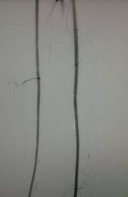
Fig 7. Follow-up angiography of the right lower leg after embolization. There is no filling of the hemangioma.
Example 4. A case of combined treatment of an arteriovenous malformation in the eye region using general vascular endovascular and neuroendovascular technologies.
Patient L., 43 years old. Complaints of a tumor-like lesion in the right eye region with signs of vascular pulsation. Angiography revealed an arteriovenous malformation (AVM) with an aneurysm in the AVM region (fig. 1). A special feature of this clinical case is that the AVM fills from the territories of both the external and the internal carotid artery (fig. 1,2)
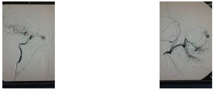
For treatment, the first stage was endovascular embolization of the AVM in the external carotid artery territory by superselective catheterization of the a. maxilaris with a 4F catheter, using general vascular endovascular technology (fig 3,4)
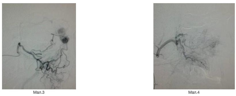
The second stage was endovascular embolization of the AVM from the internal carotid artery territory by superselective catheterization of the a. ophtalmica with a 2.5F flow-directed microcatheter, using neuroendovascular technology (fig 5, 6).

The second stage was endovascular embolization of the AVM from the internal carotid artery territory by superselective catheterization of the a. ophtalmica with a 2.5F flow-directed microcatheter, using neuroendovascular technology(fig 5, 6).
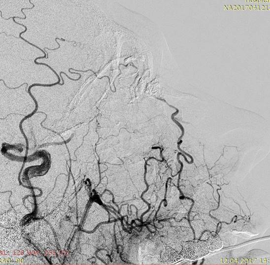 Fig.7
Example 5. AVM of the auricle. Patient K., 36 years old. Congenital arteriovenous malformation of the right auricle. Bleeding from the varicose nodes of the AVM. Condition after suturing of the AVM varices. Recurrent bleeding. Underwent elective treatment at the SI “SPCENRS of the NAMSU ” with complaints of deformity of the right auricle and discoloration of the skin of the right earlobe with large varicose dilated nodes showing signs of vascular pulsation and bruit (Fig. 1.).
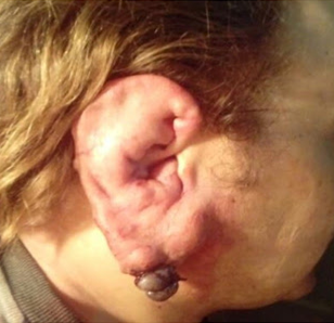
Fig. 1. Arteriovenous malformation of the right auricle.
In January 2017, profuse bleeding from the varicose nodes developed. In the ENT department of the regional hospital in Poltava, the earlobe varices were sutured. A week later the bleeding recurred. Based on CT, a diagnosis of arteriovenous malformation was made (Fig. 2.).
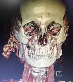
Fig. 2. CT angiography. Arteriovenous malformation of the right auricle
At the SI “SPCENRS of the NAMSU” for diagnostic purposes, angiography of the right ECA and ICA was performed on 06.03.17. Angiography revealed an arteriovenous malformation of the right ear with active blood flow and filling of the AVM from the territory of the right posterior auricular artery (Fig. 3.4.).
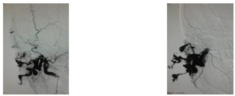
Fig. 3,4. Angiography. Arteriovenous malformation of the right auricle
For treatment, on 06.03.17 embolization of the auricular AVM in the territory of the right posterior auricular artery was performed with emboli and embolization coils. As a result of the operation, improvement was noted in the form of cessation of bleeding and disappearance of vascular pulsation in the malformation area (Fig. 5,6.)

Fig. 5,6. Embolization of the right auricle AVM