


Spinal arteriovenous malformations (AVMs) are a heterogeneous group of non-neoplastic diseases representing an abnormal plexus in which arteries are directly connected to veins through an abnormal capillary connection. An arteriovenous fistula (AVF) is a direct connection between an artery and a vein. It accounts for 3–4 % of spinal cord diseases that cause neurological deficit and lead to inevitable disability. With timely diagnosis, treatment has a positive clinical effect. When choosing a treatment method, the degree of neurological deficit and the results of spinal angiography (SAG), in particular the features of the angioarchitecture, must be taken into account. Total exclusion of the AVM from the circulation is key to preventing progression of neurological deterioration; however, the goal of treatment is functional preservation of the spinal cord blood supply [2]. In some cases, reducing flow through the shunt helps relieve symptoms . Combining endovascular and microsurgical techniques increases the effectiveness of treatment in patients with spinal AVMs.
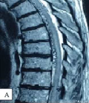
Patient with an AVM
A – MRI of the thoracic spine: dilated tortuous perimedullary vessels; B – selective spinal angiography: at the L1–L2 level, an artery feeding the arteriovenous fistula (white arrow) and arising from the left segmental artery is visualized, as well as the draining vein (black arrow); C – microsurgical disconnection of the arteriovenous fistula was performed; D – postoperative selective spinal angiography. Total exclusion of the dural fistula was achieved (black arrow).


