


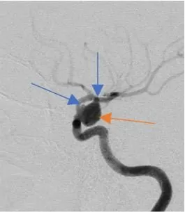
Patient: S., 27 years old
Acute onset on 28.11.2021: impaired consciousness, nausea, a single episode of vomiting. Computed tomography of the brain performed at the place of residence diagnosed a subarachnoid hemorrhage; computed angiography of the cerebral arteries showed signs of a saccular aneurysm of the supraclinoid segment of the right internal carotid artery.
The patient was admitted to the Center on day 2 of the disease. Clinical status on admission (30.11.2021): no focal neurological deficit, Hunt-Hess IІ, WFNS-II.
Diagnosis: Cerebrovascular disease: Saccular aneurysm of the supraclinoid segment of the right internal carotid artery. Acute cerebrovascular accident of the subarachnoid hemorrhage type (28.11.2021).
Selective digital subtraction cerebral angiography shows a complex-shaped saccular aneurysm of the supraclinoid segment of the right ICA with a wide neck and two diverticula. Aneurysm size – 8.9×7.3 mm, aneurysm neck 6.4 mm. Saccular aneurysm of the supraclinoid segment of the right internal carotid artery (Fig. 1, 2, 3, marked with a red arrow) with a diverticulum (rupture site, blue arrow). Rotational digital subtraction angiography (video 1).

Choice of surgical strategy: given the acute period of the hemorrhage and the wide aneurysm neck, total reconstructive and, above all, stable occlusion of the aneurysm cavity with microcoils required implantation of a protective stent at the level of the aneurysm neck. To increase the radicality of the operation, a FRED flow-diverting stent (3.5/22/16 mm) was chosen.
The patient underwent endovascular surgery on day 2 of the disease. 45 minutes before the operation the patient received BRILINTA 180 mg.
Course of the operation: The marker of the Headway17 microcatheter (Microvention Terumo) was advanced into the aneurysm cavity; the stent delivery microcatheter was placed distal to the aneurysm (Fig. 7). With the stent partially deployed at the level of the aneurysm neck, framing (forming a frame inside the aneurysm cavity) was performed with an AXIUM™ PRIME FRAME coil (Medtronic). The aneurysm cavity was filled (filling) with 4 ED COIL∞10 Soft microcoils (Kaneka) until a coil conglomerate of metallic density was formed (Fig. 8, 9). After stent implantation, follow-up angiography demonstrates total reconstructive (with preserved patency of the parent artery). (Fig 10, 11. Arrows indicate the markers of the implanted stent).


Follow-up angiography at the end of the operation showed mechanical vasospasm of the M1 segment of the middle cerebral artery and of the supraclinoid segment of the carotid artery at the level of the stent markers (Fig. 12).

Balloon angioplasty of the spastic arterial segments was performed using an Occlusion balloon Catheter – Scepter XC (Fig. 13 – 16. Arrows indicate the balloon at the level of the spastic arterial segments)

After angioplasty – restoration of arterial patency (Fig. 17,).

Brain CT immediately after the operation (Fig 18, 19). The extent of the hemorrhage corresponds to the initial computed tomography.

01.12.2021. Follow-up cerebral angiography one day after the operation: Angiographically – no vasospasm, the stent is patent with no signs of thrombi in the lumen. Normalized anatomy of the supraclinoid segment of the right ICA (Fig. 20 – 22).

Cerebral angiography (follow-up) figure 35.
The patient was discharged 2 weeks after the operation in satisfactory condition, without focal neurological deficit, GCS – 15 points (Video 4). The patient’s consent to publication of the photos has been obtained.


