


Patient S., 22 years old, was referred to the SI “SPCENRS of the NAMSU” for stenosis of a transplanted renal artery.
History: the illness began in early 2024. He had acute glomerulonephritis of both kidneys, followed by impaired function of both kidneys and the development of renal failure. Despite regular courses of hemodialysis for six months, renal failure progressed to failure of both kidneys.
In August 2024, at the National Children’s Specialized Hospital “Okhmatdyt”, a donor kidney was transplanted with the renal artery anastomosed to the right internal iliac artery. The operation was technically and clinically successful.
However, at follow-up three months after transplantation, CT angiography revealed stenosis of the transplanted renal artery in the anastomosis zone. According to ultrasound, the blood flow velocity in the stenosis zone increased from 150 ml/sec to 300 ml/sec (Fig 1).
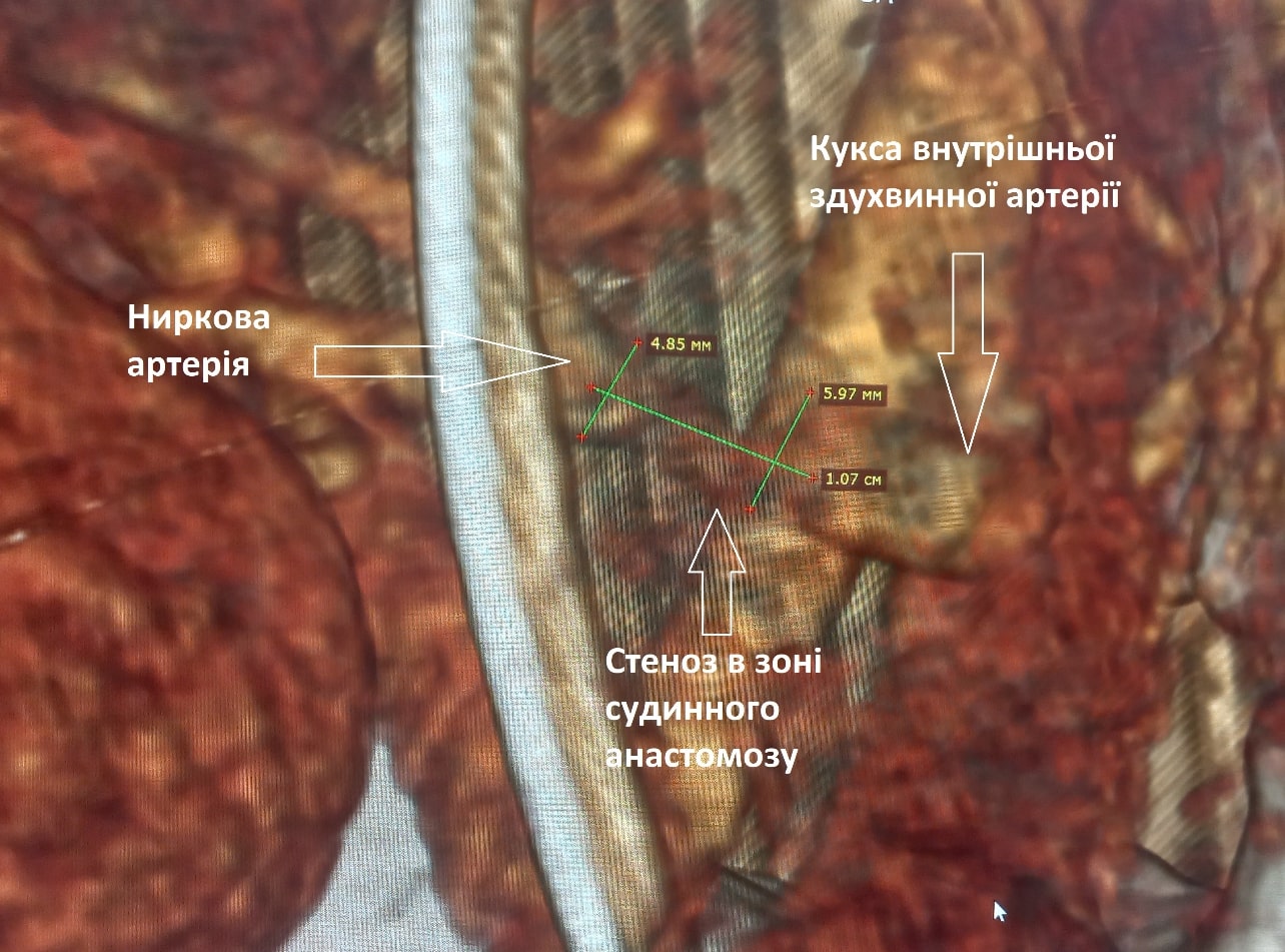
Figure 1. Stenosis of the transplanted renal artery in the vascular anastomosis zone on CT angiography
For diagnostic purposes, on 03.10.24 the patient underwent arteriography of the transplanted kidney via right femoral access. The angiogram shows the renal artery of the transplanted kidney arising from the stump of the right internal iliac artery. There is 80-90 % stenosis of the renal artery in the middle third, 1.0 cm long, in the vascular anastomosis zone. The renal artery arises from the common iliac artery at an acute angle, which makes its catheterization via right femoral access impossible (Fig 2).
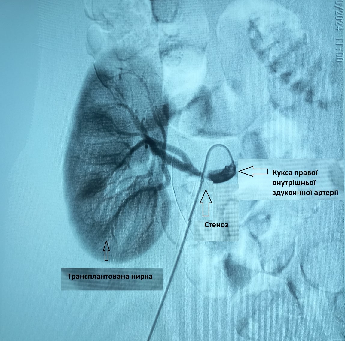
Figure 2. Arteriography of the transplanted kidney from the right common femoral artery. Stenosis of the transplanted renal artery in the vascular anastomosis zone
A decision was made to stent the renal artery via contralateral access through the left common femoral artery.
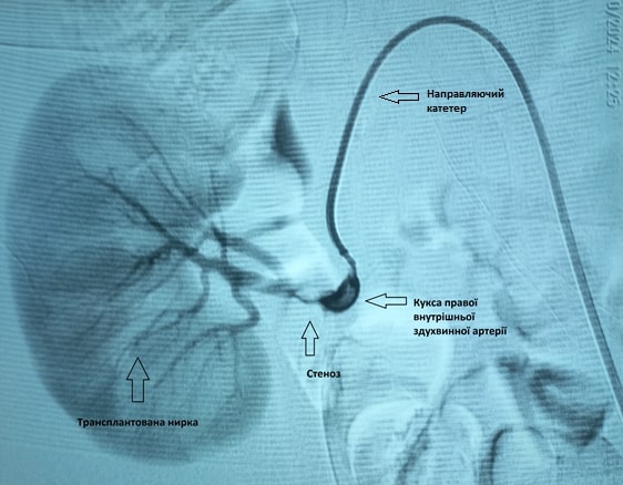
Figure 3. Contralateral catheterization of the right internal iliac artery from the left common femoral artery. Stenosis of the transplanted renal artery in the vascular anastomosis zone
Under local anesthesia, Sol Lidocaini 0.5% – 20.0 ml, the left common femoral artery (CFA) was punctured using the Seldinger technique. A 6F introducer was placed in the CFA. Through the introducer, over a guidewire, using a guiding catheter, the right internal iliac artery and the transplanted renal artery were catheterized contralaterally under fluoroscopic control (Fig 3). Next, a microguidewire was advanced through the guiding catheter into the renal artery and passed beyond the stenosis zone. Then, through the guiding catheter over the microguidewire, a balloon-mounted stent 5.0 mm in diameter and 19 mm long was advanced into the stenosis zone of the renal artery (Fig 4).
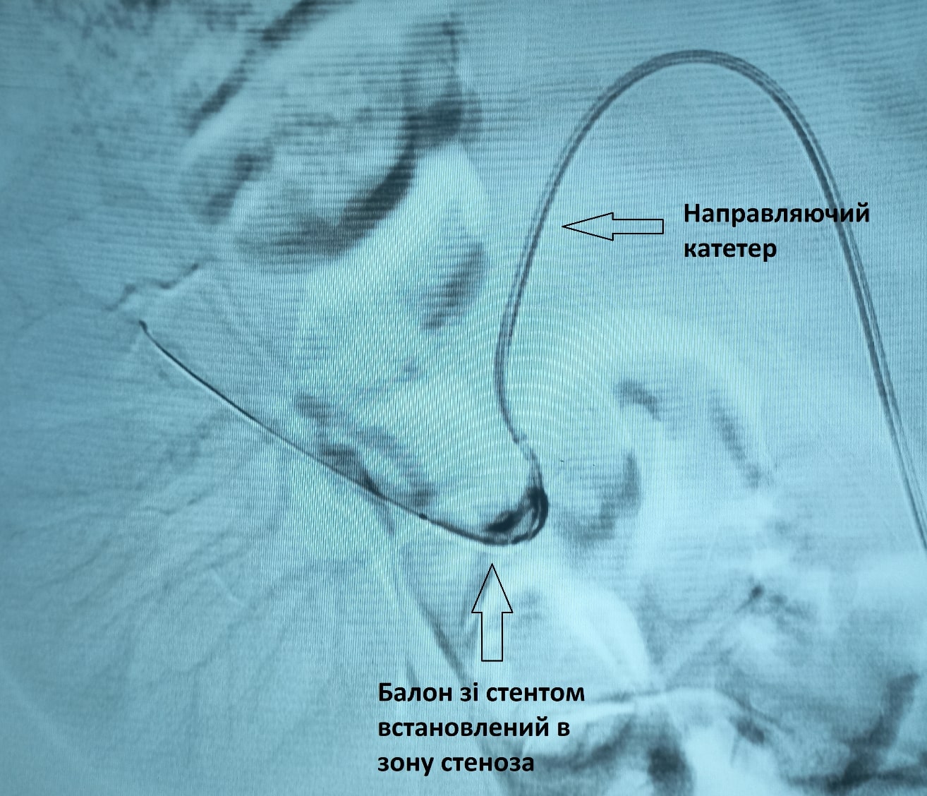
Figure 4. A balloon-mounted stent 5.0 mm in diameter and 19 mm long was advanced through the guiding catheter over the microguidewire into the stenosis zone of the renal artery
Stenting of the transplanted renal artery stenosis was performed. The control angiogram shows no renal artery stenosis. Patency of the transplanted renal artery was fully restored (Fig 5,6).
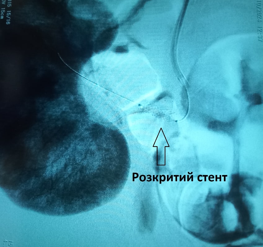
Figure 5. The stent is deployed in the stenosis zone of the transplanted renal artery.
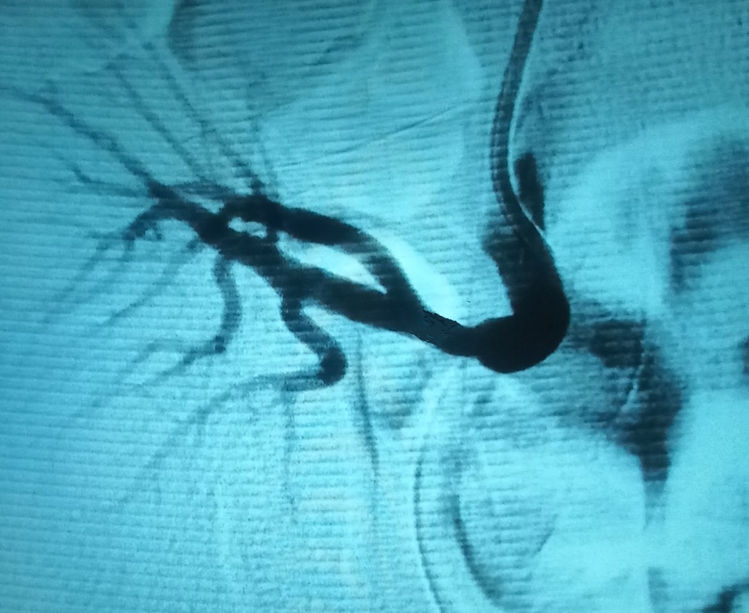
Figure 6. Control angiography. The control angiogram shows no renal artery stenosis. Patency of the transplanted renal artery was fully restored
The postoperative period was uneventful. The next day the patient was transferred to the National Children’s Specialized Hospital “Okhmatdyt” in satisfactory condition.


