


Patient: L., 61 years old.
She has considered herself ill since 2019, when she began to suffer from periodic headaches and unstable blood pressure, which led to MSCT angiography on referral from her attending neuropathologist.
The patient was admitted to the Center electively in satisfactory condition. Neurological status without pathological findings, except for end-point nystagmus to the left.
Diagnosis: Cerebrovascular disease: Saccular aneurysm of the basilar artery apex.
Selective digital subtraction cerebral angiography determined the X-ray angiographic characteristics of the aneurysm and the surrounding arteries: a saccular aneurysm (SA) of the apex of the basilar artery (BA) with a wide neck, extending to the proximal segment of the left posterior cerebral artery. Aneurysm size – 4.9×4.8 mm, aneurysm neck width 4.7 mm (Fig. 1, 2, 3).

Fig. 1. Saccular aneurysm of the basilar artery apex.
SA – saccular aneurysm; PCA – posterior cerebral artery; BA – basilar artery
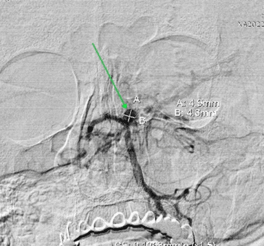
Fig. 2. Dimensions of the saccular aneurysm of the basilar artery apex.
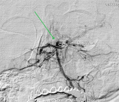
Fig. 3. Measurement of the neck width of the BA SA
Choice of surgical strategy. For reconstructive total exclusion of the saccular aneurysm from the circulation in order to prevent its rupture with a hemorrhagic stroke, and taking into account the location of the aneurysm (the basilar artery apex at its bifurcation into the right and left posterior cerebral arteries) and the wide neck, it was decided to occlude the aneurysm cavity with microcoils using a pCONUS 2 bifurcation implant (4mm*15mm*6mm* ID 0.021 V2,5-3,7).
The patient underwent elective surgery after preoperative preparation with dual antiplatelet therapy: Brilinta 90 mg* 2 times a day + ASA 100 mg * 1 time a day.
Course of the operation: Phenom 0,021 (Medtronic) and Headway 0,017 (Microvention Terumo) microcatheters were advanced over microguidewires to the level of the saccular aneurysm of the basilar artery (Fig.4).
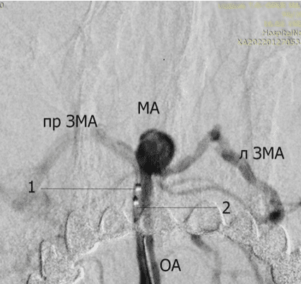
Fig. 4. Placement of the microcatheters in the distal segment of the basilar artery (BA).
1 – Phenom 0,021 microcatheter (Medtronic)
2 – Headway 0,017 (Microvention Terumo)
The Phenom microcatheter for pCONUS 2 implantation was advanced and positioned in the aneurysm cavity (Fig.5).
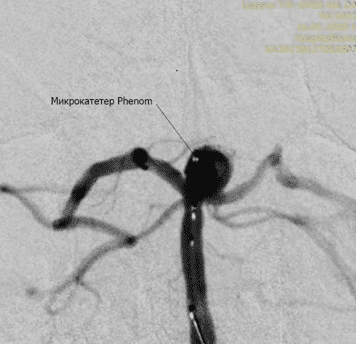
Fig. 5. The Phenom microcatheter advanced into the cavity of the basilar artery SA
The microguidewire was removed. The pCONUS 2 bifurcation implant was advanced through the Phenom microcatheter and deployed so that the petals of the crown lay intrasaccularly in the aneurysm cavity, creating a stabilizing frame in the area of its neck. The shaft of the implant lay in the basilar artery, closely adhering to its walls, stabilizing the implant without impairing blood flow in the artery (Fig.6).
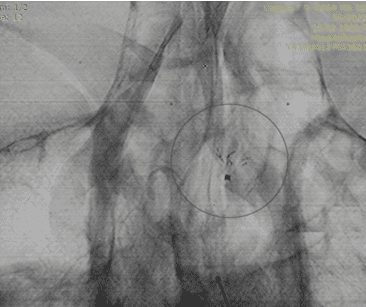
Fig. 6. The pCONUS 2 bifurcation implant is deployed.
The crown with the petals is in the aneurysm cavity.
The shaft of the implant is in the basilar artery.
The Headway 0,017 microcatheter was advanced over a microguidewire through pCONUS 2 into the cavity of the saccular aneurysm and positioned without touching the aneurysm walls (Fig.7, 8).
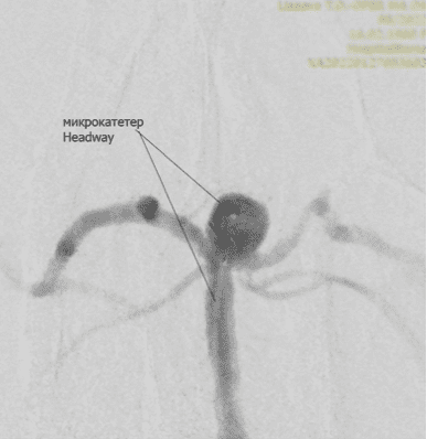
Fig. 7. The Headway 0,017 microcatheter is positioned in the cavity of the basilar artery SA
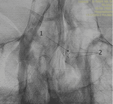
Fig. 8.
1 – Marker of the Headway microcatheter positioned in the SA cavity;
2- petals of the pCONUS 2 implant crown.
The microguidewire was removed, and MicroPlex ® 3D-configuration platinum microcoils sized 5 mm×22 cm; 4 mm×12 cm; 4 mm×12 cm; 2,5 mm×4 cm; 2 mm×2 cm (Microvention Terumo) were deployed one after another through this microcatheter until the cavity of the saccular aneurysm was completely filled and excluded from the circulation (Fig.9).
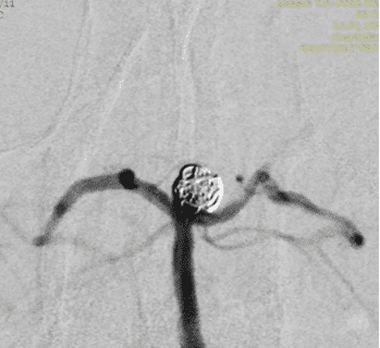
Fig. 9. Occlusion of the SA cavity with microcoils.
Subtraction angiography was performed during microcoil implantation. After the endovascular microconstruction was formed, follow-up angiography demonstrated reconstructive exclusion of the saccular aneurysm from the circulation. There were no extravasations, ischemic or other complications. Patency of all cerebral arteries is preserved (Fig. 10).
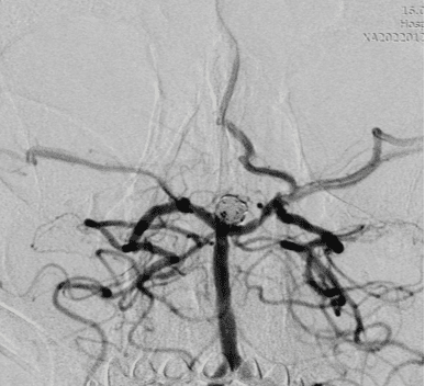
Fig. 10. Final stage of the operation – follow-up angiography.
The diagram of the endovascular microconstruction is shown below, where BA is the basilar artery, at whose apex lies the saccular aneurysm excluded from the circulation with microcoils; the aneurysm base has protective stabilization by the petals of the pCONUS 2 crown. The pCONUS 2 shaft lies in the basilar artery. PCA L(R) – left and right posterior cerebral arteries. VA L (R) – left and right vertebral arteries (Fig. 11).
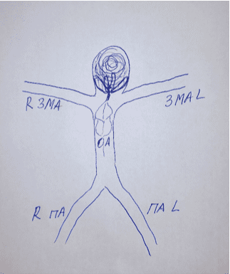
Fig. 11.
Diagram of the endovascular microconstruction
for exclusion of the BA apex SA
from the circulation with microcoils with pCONUS 2 assistance.
The patient was discharged on the second day after the operation in satisfactory condition, without pathological neurological symptoms.


